Interesting Case of the Month - (IGHP, ICOM) - April
april 2026
Clinical History-
82-year-old female complaint of abdominal pain. USG whole abdomen showed a lesion in liver. Serum AFP: 3.1 ng/mL; CA19-9: 2 U/mL.
From the images below identify the likely disease?
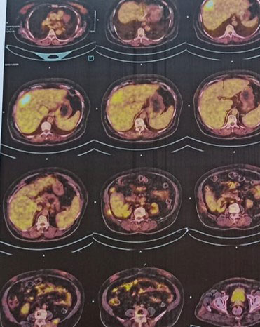
Radiology-
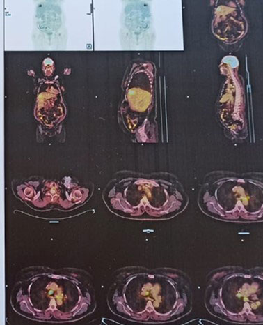
PET-CT revealed an FDG avid (SUV max 10.3), poorly circumscribed heterogeneous mass lesion measuring approximately 4.0 × 5.4 cm in the subcapsular region of segment VIII of liver with associated intralesional calcific foci. Background liver showed features of chronic liver disease with fatty change.
MRI Upper Abdomen: Mixed signal intensity lesion in segment VIII of liver which shows T2 hypointense component and T2 intermediate soft tissue signal intensity component.
Microscopic Images-
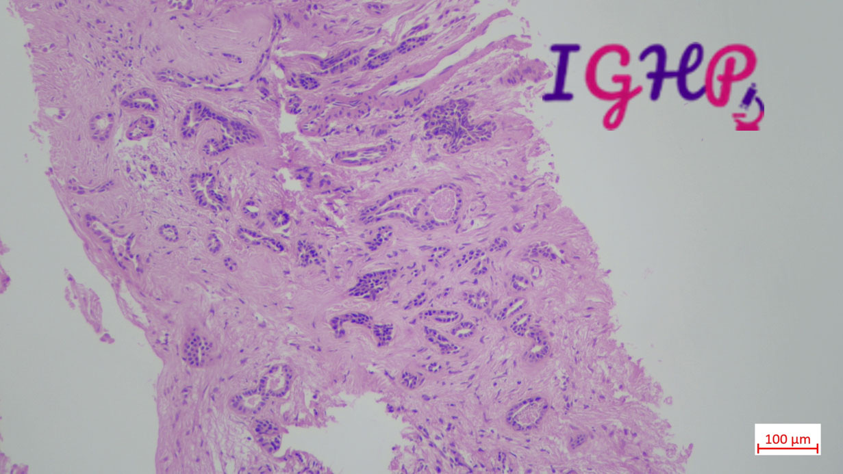
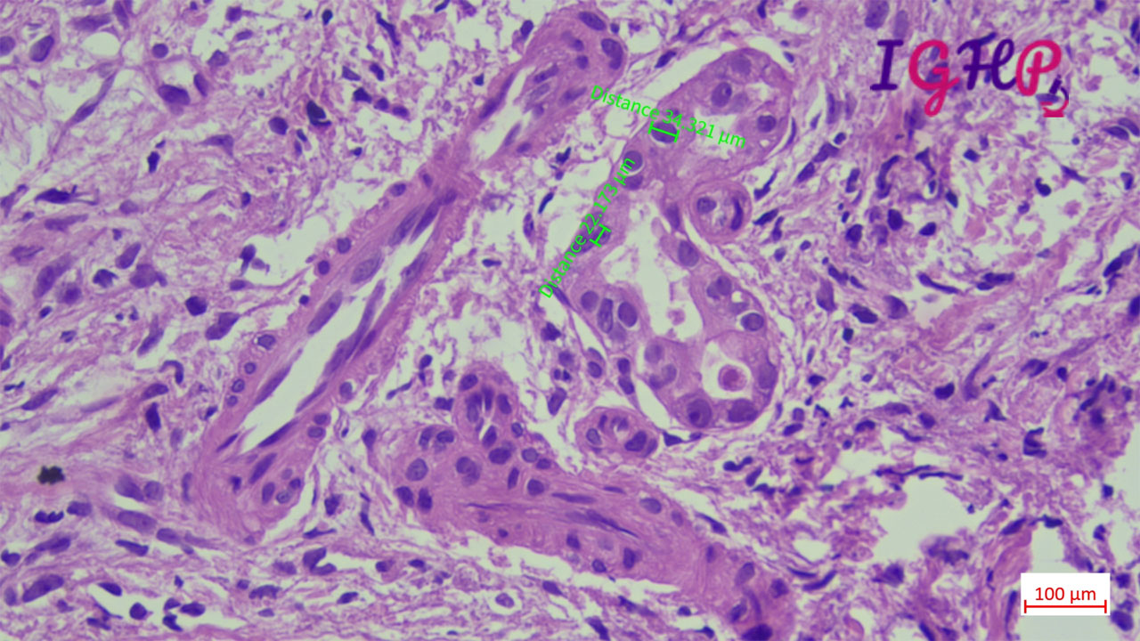
Microscopic examination showed atypical bile ductular proliferation in dense sclerotic stroma. Areas of mild nuclear pleomorphism noted. Intraluminal apoptotic debris seen.
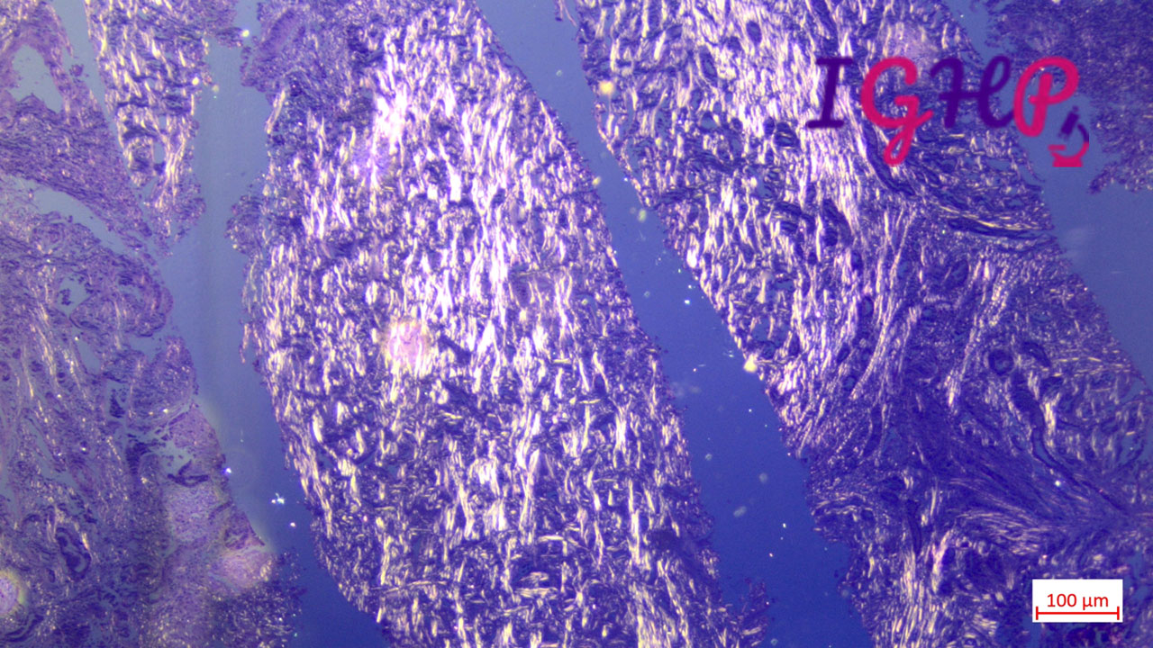
Special Stains Performed-
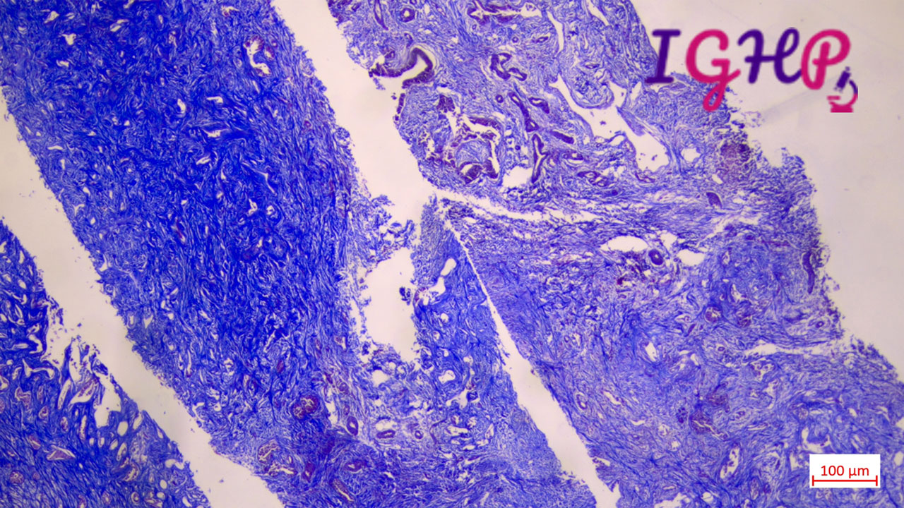
MT stain performed showed dense sclerotic desmoplastic stroma
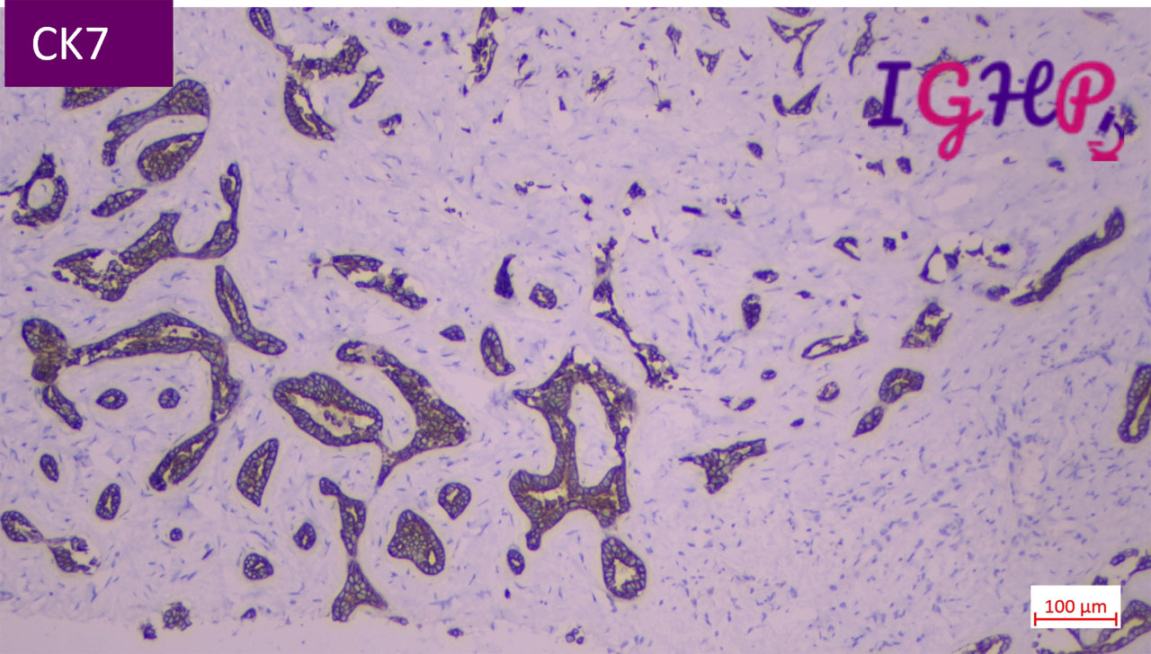
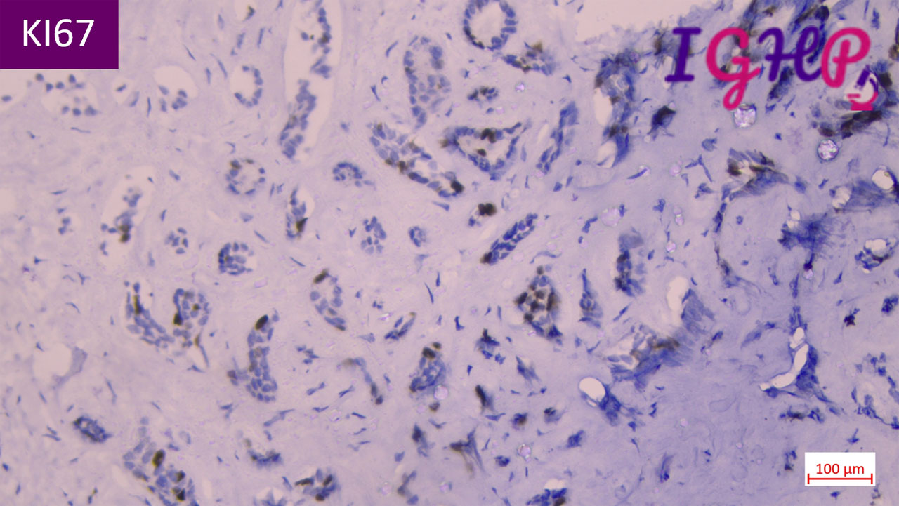
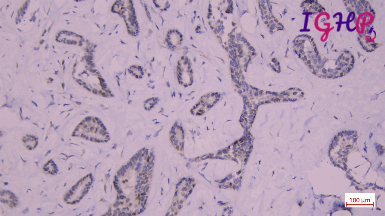
Immunohistochemistry Performed-
IHC performed for CK7, KI67 and P53