Interesting Case of the Month - (IGHP, ICOM) - October
october 2025
Clinical History-
64-year-old male history of road traffic accident presented to emergency department with severe pain abdomen. Plain CT showed ruptured mass in upper pole of kidney. From the images below identify the likely disease.
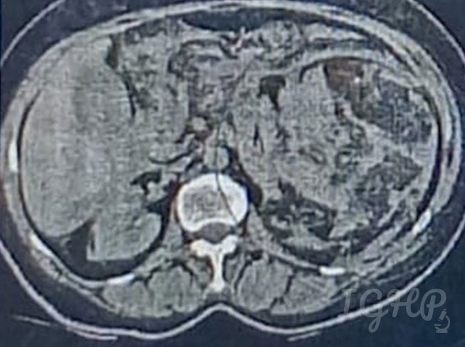
Radiology-
Plain CT-Large heterogenous exophytic space occupying mass lesion noted arising from upper pole of kidney measuring 11.4 x 10 showing fat attenuation with multiple irregular haemorrhage.
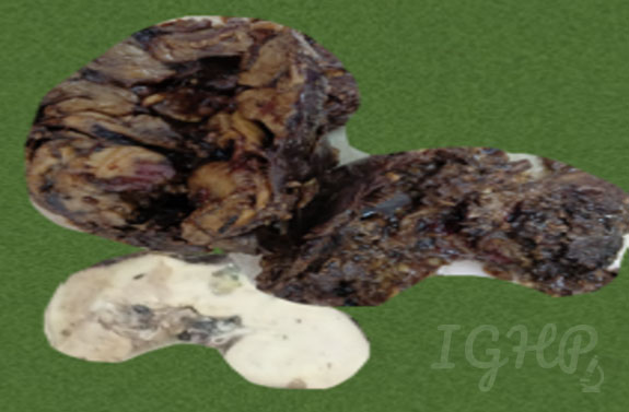
Gross Images-
The kidney upper pole showed a large, irregular, hemorrhagic mass measuring approximately 18 x 12 x 6 cm . The cut surface of the mass reveals a heterogeneous appearance with areas of haemorrhage, necrosis, and yellowish-tan tissue. The remaining renal parenchyma showed a small multi-cystic lesion noted in renal cortex of mid pole in pericapsular region.
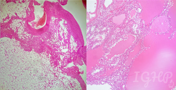
Microscopic Images-
Microscopic examination from the large mass revealed a classic triphasic pattern consisting of mature adipose tissue, myoid spindle cells, and several thickened and dysmorphic blood vessels of varying sizes. Microscopy from the small discoloured cystic area in middle pole showed features of multilocular clear cell renal cell neoplasm of low malignant potential.