Interesting Case of the Month - (IGHP, ICOM) - May
may 2026
Clinical History-
22-year-old femalecomplaint of abdominal pain. H/o Left lobe liver cyst with recurrent cyst infections, cholangitis? Caroli’s disease. From the images below identify the likely disease?
Radiology-
CT mild central and peripheral intrahepatic biliary nodule dilatation. Mild hypodense non enhancing lesions in both liver lobes.
Microscopic Images
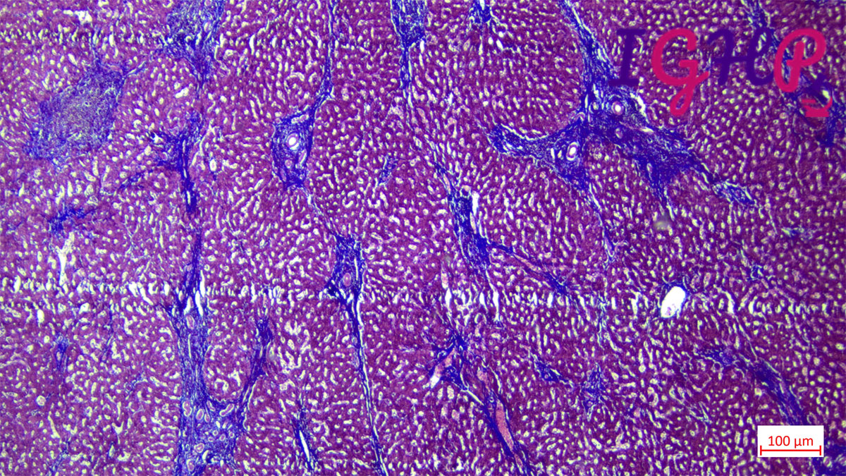
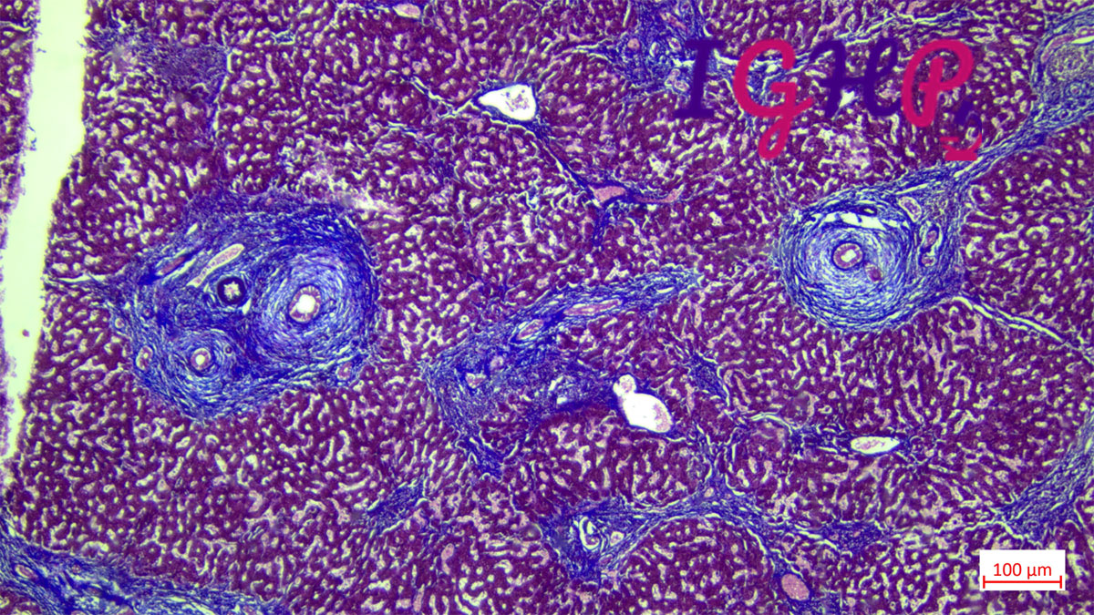
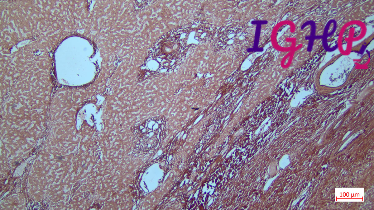
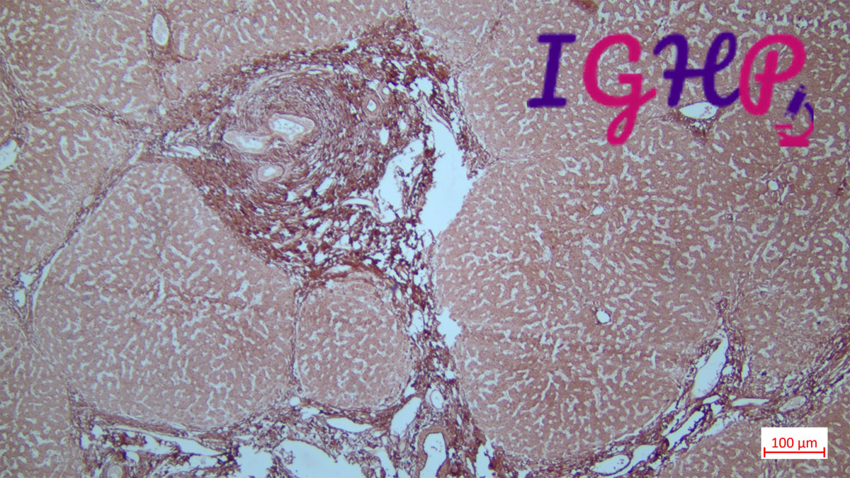
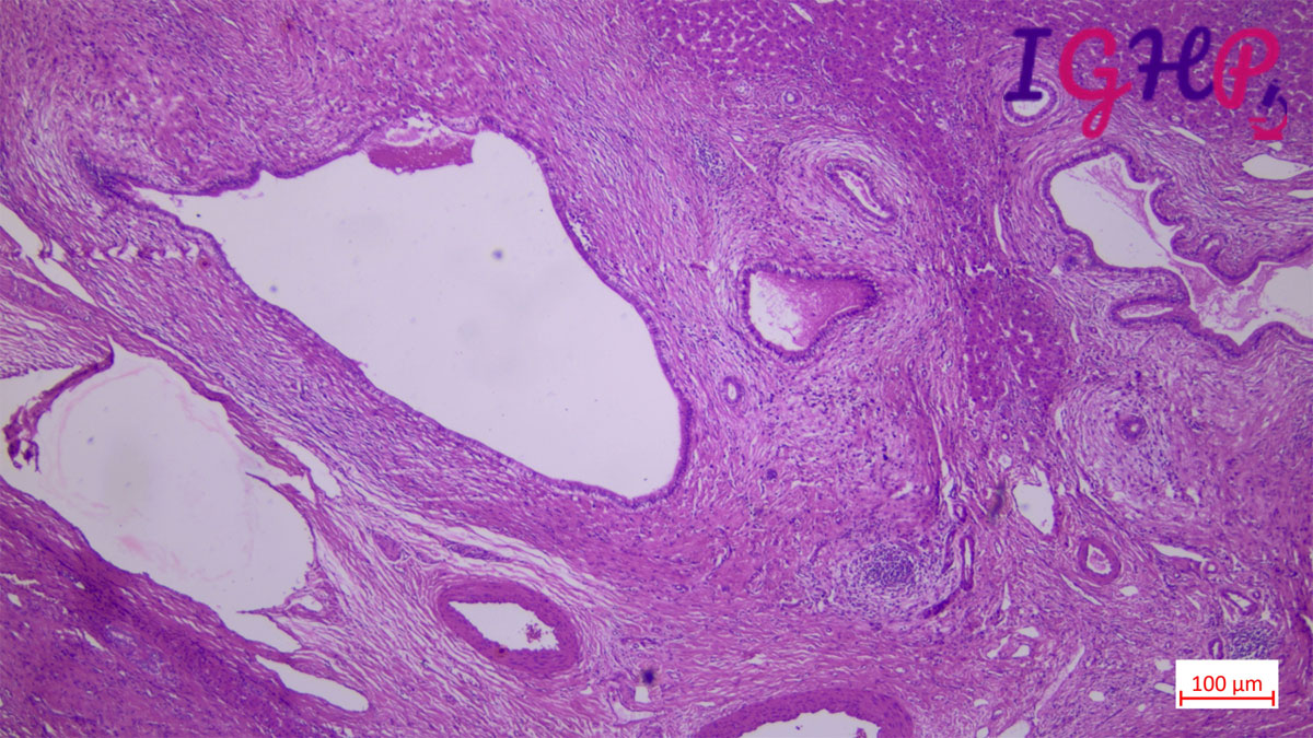
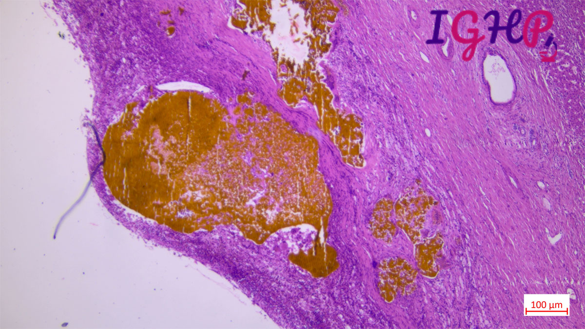
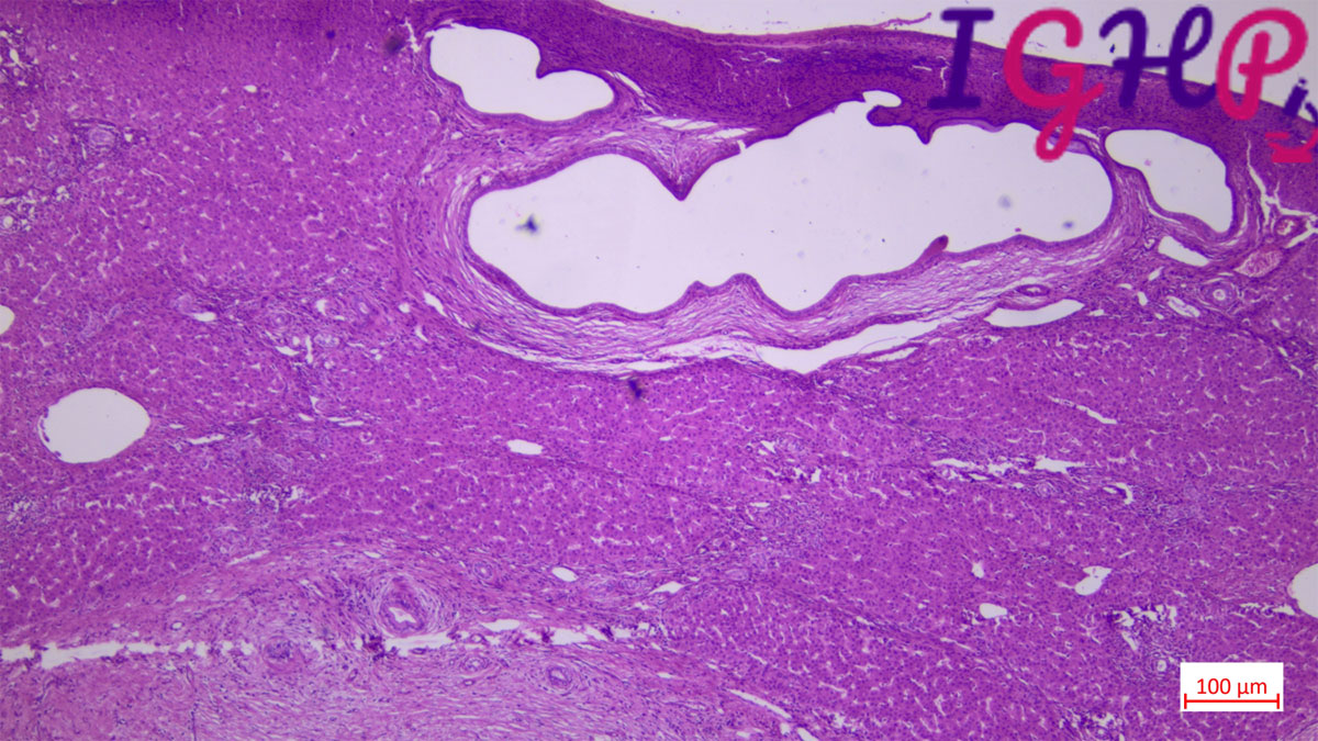
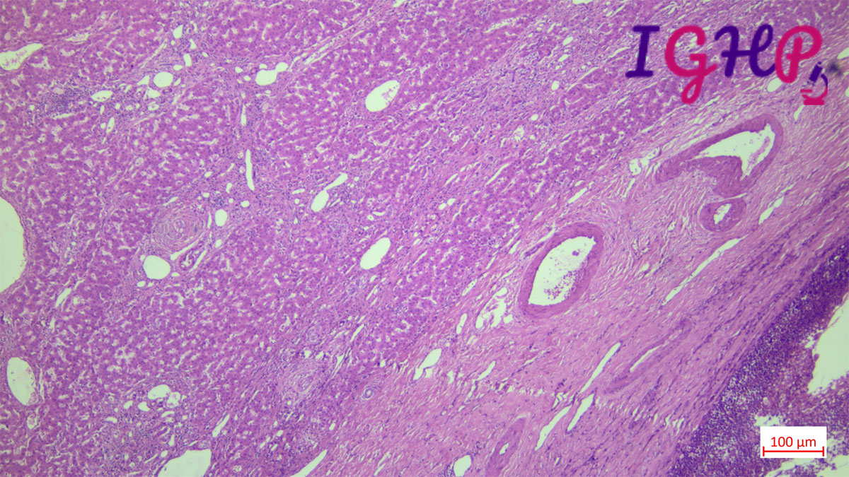
Microscopic examination of the liver sections demonstrates multiple, large, saccular dilatations of the intrahepatic bile ducts. The background liver parenchyma show areas of fibrous cholangitis with presence of a mixed inflammatory infiltrate, either within, or adjacent to, damaged bile ducts associated with extensive collagen deposition. There are presence of bile plugs within the bile ducts and the hepatic arteries appear hyperplastic.
Special Stains Performed-
MT and Orcein stain performed showed bridging fibrosis with areas of periductal concentric onion skin fibrosis