Interesting Case of the Month - (IGHP, ICOM) - March
march 2026
Clinical History-
33-year-old male with bleeding PR underwent colonoscopy which revealed a polyp on colon, EMR polypectomy was performed.
From the images below identify the likely disease?
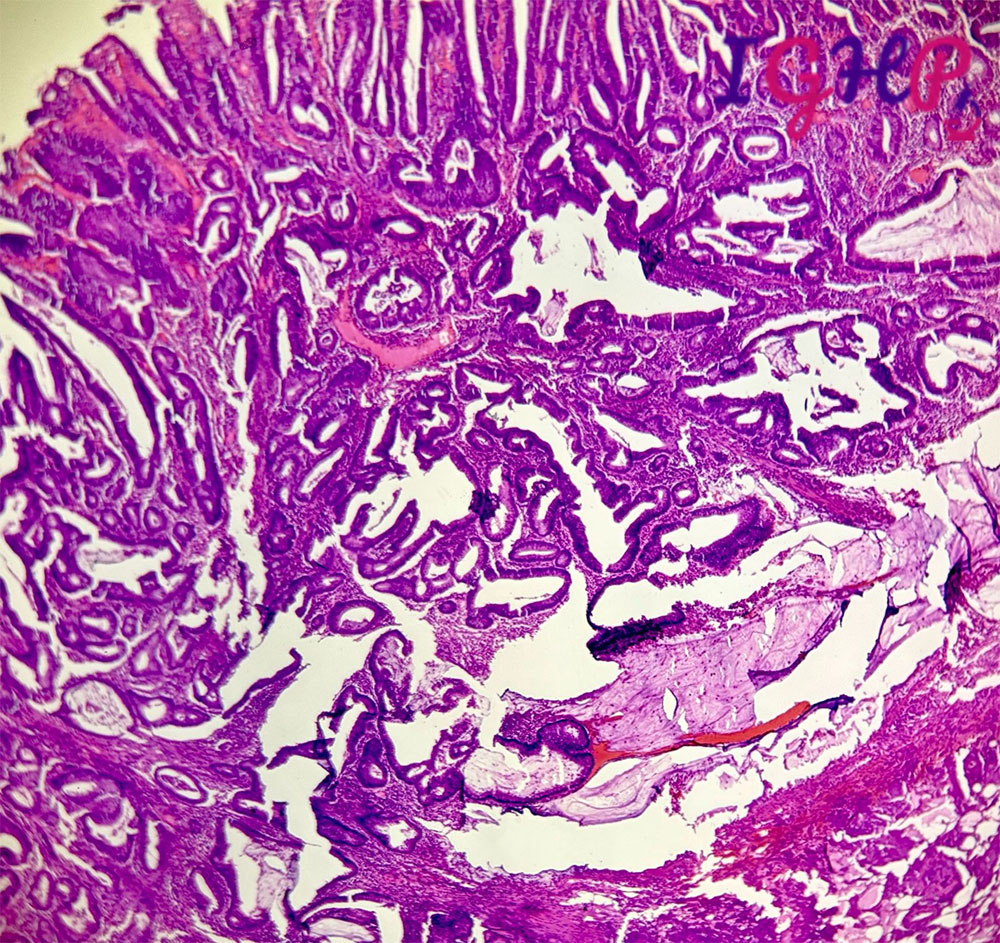
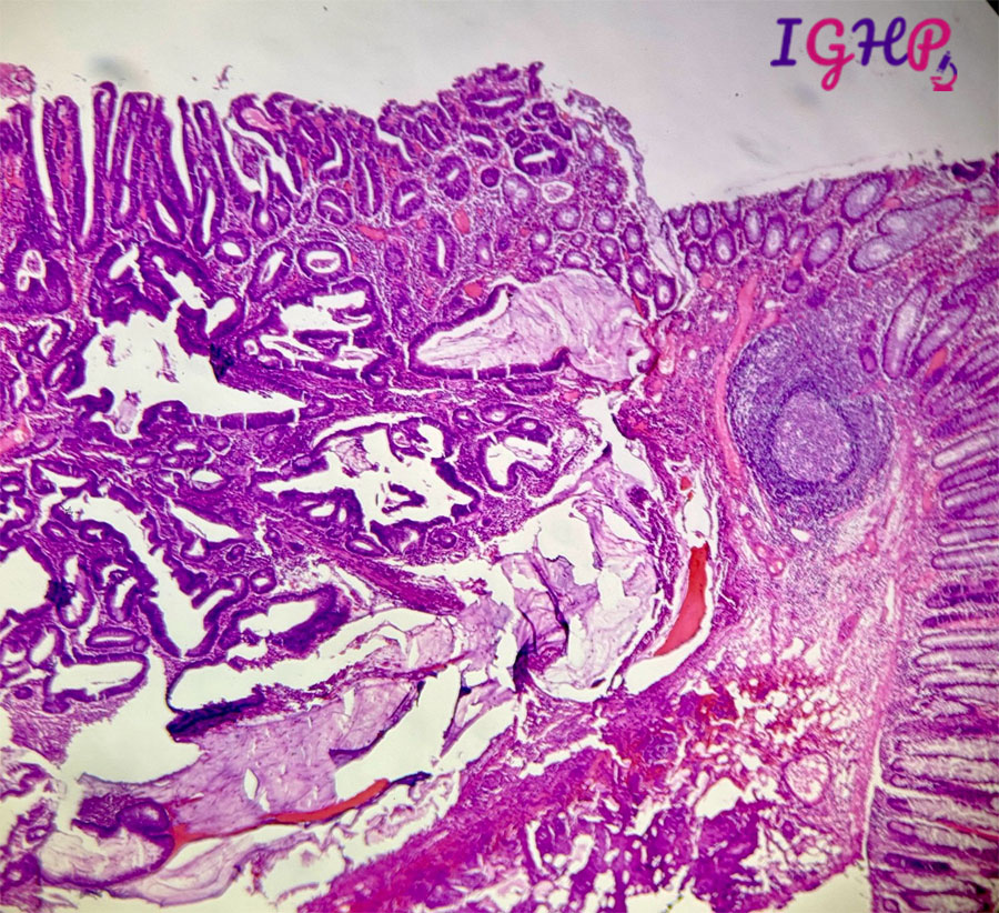
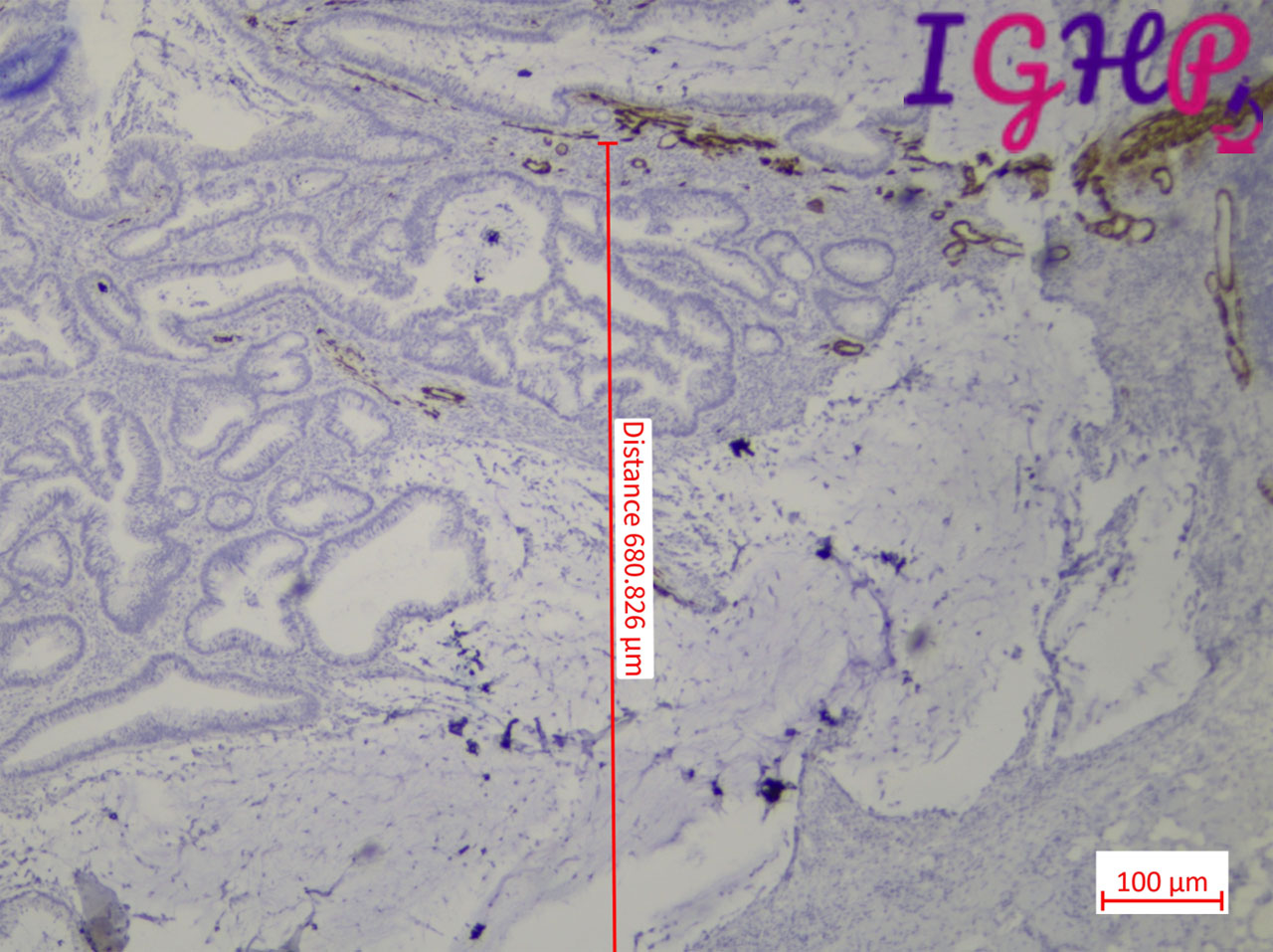
Microscopic Images-
Microscopic examination from the EMR specimen showed changes of tubular adenoma on the surface with focal area of progression into the submucosal region. The nuclear changes are focally high grade. The lesion demonstrated progression into the submucosal region. The submucosal tumor nests consist of variably sized, irregular and dilated glands showing features of atypical epithelial lining, and associated extravasated mucin.
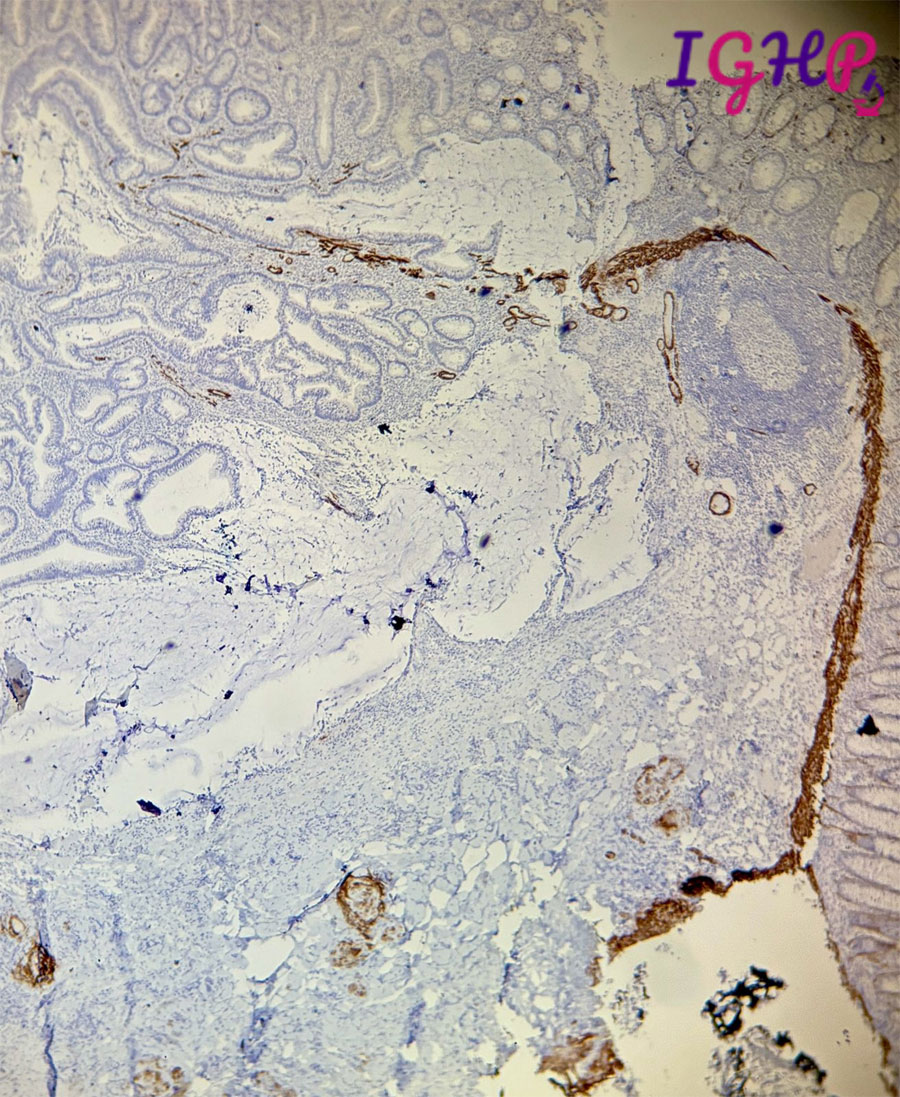
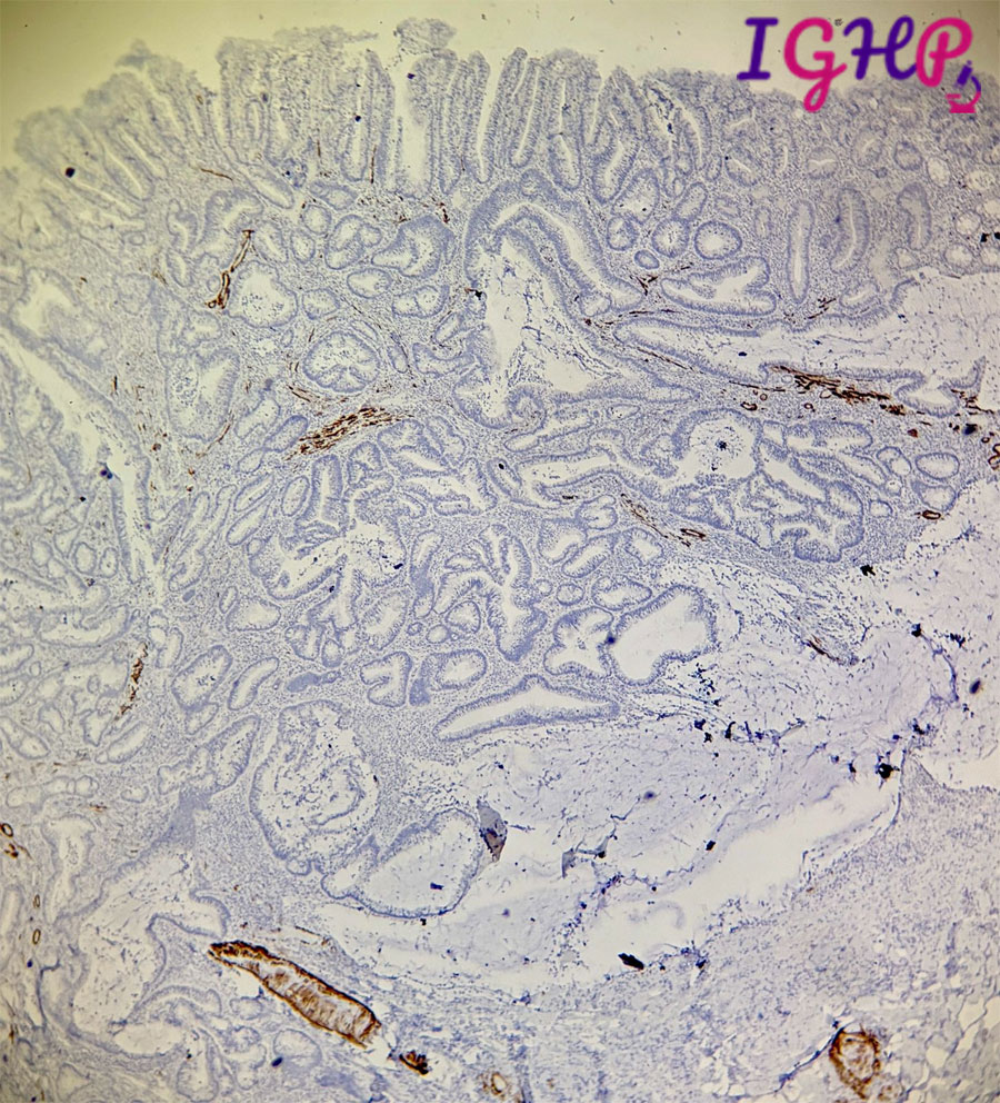
Immunohistochemistry -
Immunohistochemistry for Desmin and SMA showed presence of malignant glands within the submucosa
Diagnosis-
Tubular Adenoma, Low Grade Dysplasia
Adenocarcinoma arising in Tubular Adenoma
Tubular Adenoma, High Grade Dysplasia
Villous Adenoma, Low Grade Dysplasia


